Catapult Pharmacology
Drug-Receptor Interactions
Drug-Receptor Interactions
G-protein Coupled Receptors
A G-protein coupled receptor (Figure 1) is a 7-transmembrane protein receptor that is coupled with a G-protein (protein that binds with GTP). G-proteins have 3 subunits (α,β,γ). There are four main types of G proteins: Gαs, Gαi, Gq and G11.
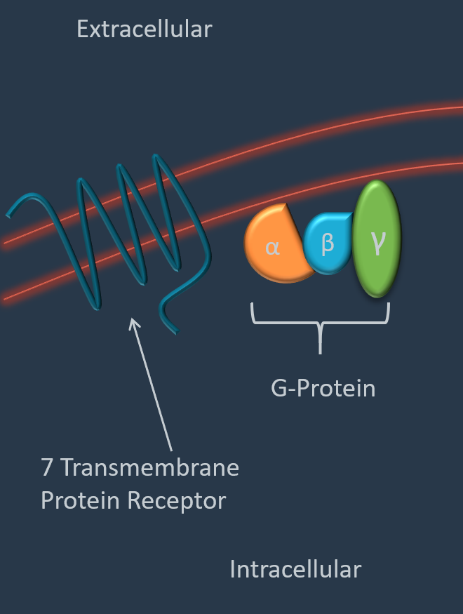
Figure 1 - G-protein coupled receptor diagram
When a drug/hormone interacts with the G-protein coupled receptor, the receptor changes conformation and interacts with the G-protein, which in turn, causes downstream signaling effects.
G-protein subtypes:
Stimulatory (Gαs)
Gαs Increases cyclic AMP (cAMP) production.
cAMP is a secondary messenger that is involved in signaling pathways such as the response to epinephrine and to vasopressin mediated water retention.
cAMP activates protein kinase A (PKA), which phosphorylates effector proteins.
The diagram below (Figure 2) shows the signaling cascade that results from activation of the Gαs protein, which results in cAMP production:
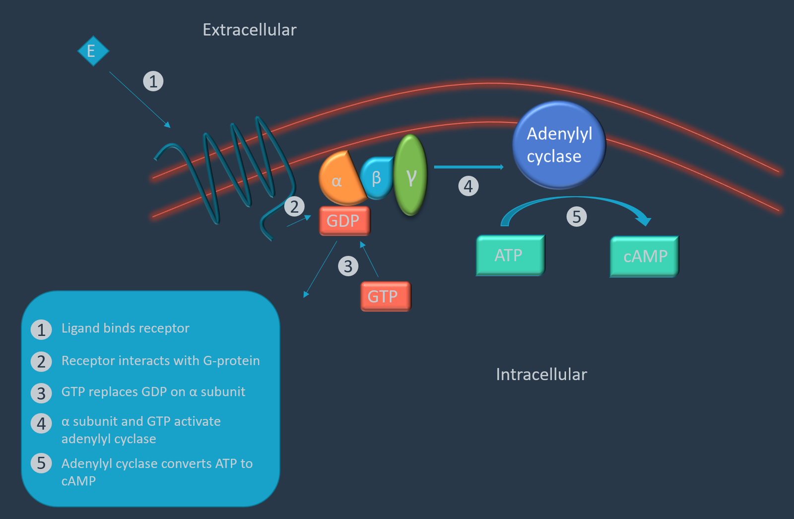
Figure 2 - Gαs Diagram
Inhibitory (Gαi)
- Gαi inhibits cAMP production, which leads to decreased levels of cAMP and PKA.
Gq and G11
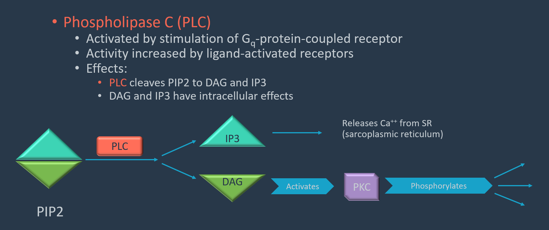
Gq and G11 increase activity of phospholipase 3 (PLC)
PLC cleaves PIP2 into DAG (diacylglycerol) and IP3 (inositol triphosphate). IP3 releases Calcium from the sarcoplasmic reticulum.
DAG activates PKC, which phosphorylates and activates other effector proteins.
Enzyme-linked Receptors (Most commonly Tyrosine Kinase Receptors)
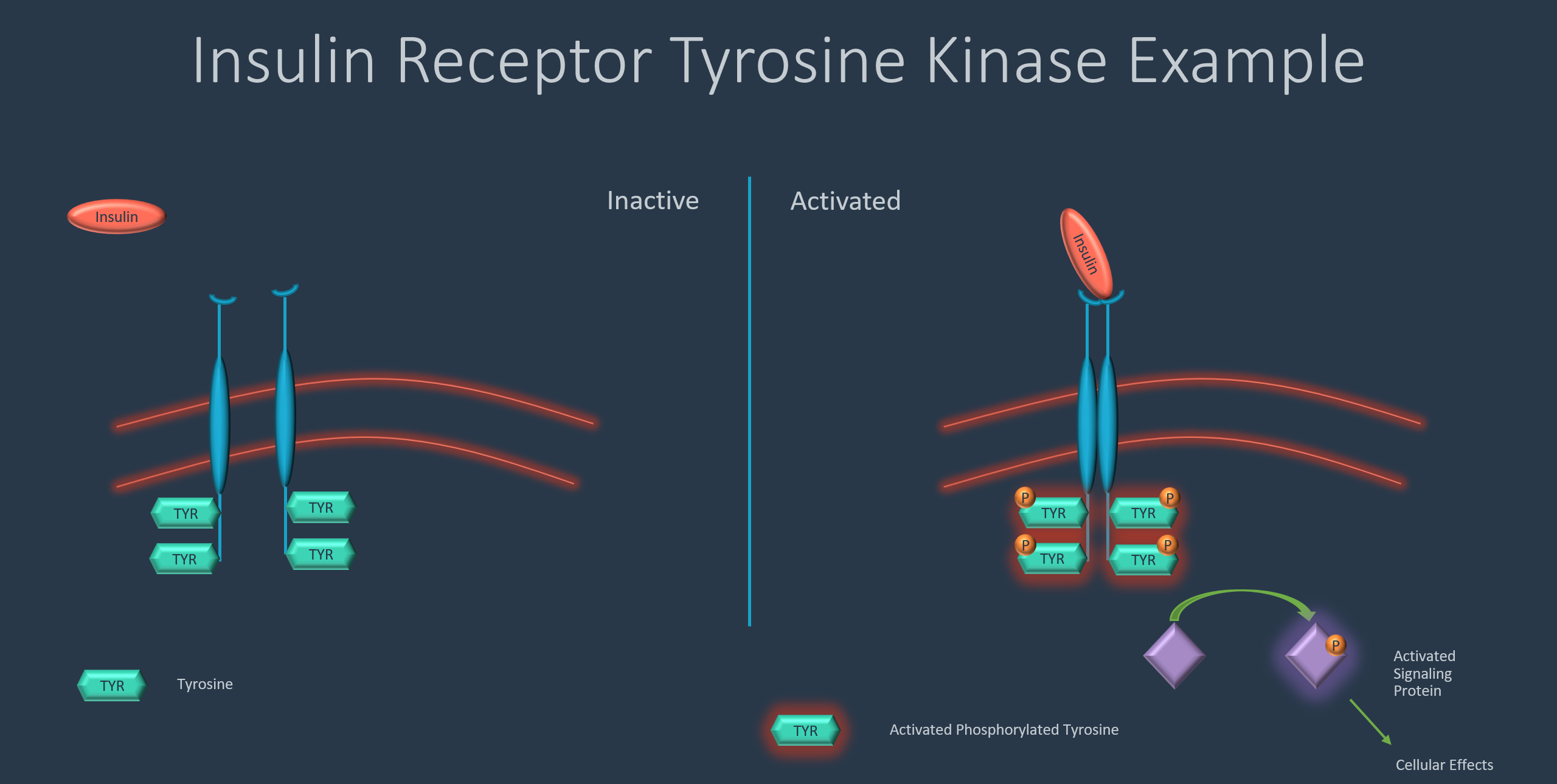
Enzyme-linked receptors are a group of receptors that form dimers or multi-unit complexes when activated by a ligand. The change in conformation results in activation of enzymes on the cytoplasmic side of the membrane. In the case of tyrosine kinase receptors, the intracellular portion of the receptor auto-phosphorylates when the ligand binds, and the conformation change occurs. The activated cytoplasmic portion of the receptor then activates other signaling proteins, which lead to their effects. Insulin and PDGF (platelet derived growth factor) use this signaling method (among other compounds).
Ligand Activated Ion Channels
Ligand activated ion channels are activated when a ligand binds an extracellular receptor. The ion channel then changes conformation and allows passage of ions through the channel.
Intracellular (intranuclear) Receptors
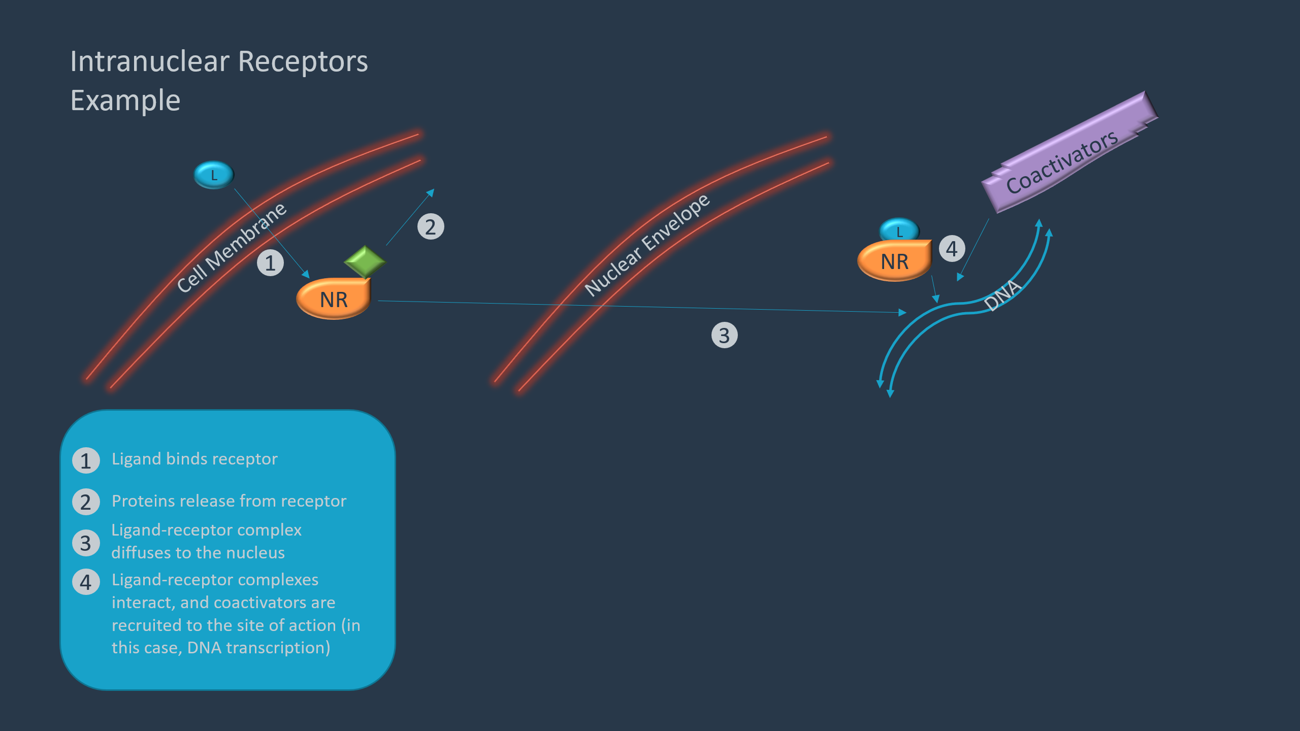
Intracellular receptors are primarily made up of intranuclear receptors that activate transcription factors. As the receptors are intracellular, the ligand must be able to pass through the cell membrane (and intranuclear envelope if a nuclear transcription factor) and thus must be lipophilic.
Lipophilic ligands, such as cortisol, pass through the cell membrane and interact with nuclear receptors in the cytoplasm. Prior to activation, the nuclear receptors are usually bound to proteins, which are released. The nuclear receptor then can translocate to the nucleus, where the activated nuclear receptor interacts with other nuclear receptors and coactivators, which increases transcription.
Other intracellular receptors interact with other intracellular contents such as proteins, enzymes, or mRNA. For example, “statin” drugs (HMG-CoA Reductase inhibitors) function by interacting with an enzyme (HMG-CoA Reductase) to decrease cholesterol production.
Antimetabolites
Antimetabolites are chemicals that are like naturally occurring compounds. Antimetabolites work by disrupting the normal cellular function. For example, 5-Fluorouracil is an analog of uracil, which is used in the production of dTMP. dTMP is a building block of DNA. When 5-fluorouracil is incorporated in the production of dTMP instead of uracil, thymidylate synthetase is inhibited, which halts DNA production.